48
@
U S T . H K
Subcellular structures can now be visualized
by super-resolution optical microscopes, to
the nanometer level.
At HKUST Super-Resolution Imaging
Center, physicists Prof Michael Loy and
Prof Shengwang Du have taken the lead to
work with life scientists, chemists, computer
scientists and mathematicians to build
state-of-the-art super-resolution localization
fluorescence microscopes that are able to
resolve tiny structures in cells or tissues
that cannot be visualized with traditional
optical microscopes. The successful
implementation of this technology now
underpins the University’s strengths in many
frontier research areas, including the study
of neurodegenerative diseases.
In an on-going study to reveal the
molecular organization of subcellular
organelles, the central research platform
developed focuses on two state-of-the-
art microscopes: one stochastic optical
reconstruction microscopy (STORM)
machine, capable of spatial resolutions
of 20 nanometers; and a light sheet
microscope with improved sample
preservation and fast acquisition times.
These advanced tools are helping scientists
explore the dynamics of synaptic vesicles
in nerve cells and lipid droplets in intestinal
cells, and the response of mitochondria
to various biological stressors, among
others. The project has received almost
HK$8 million in funding from Hong Kong
Research Grants Council.
The super-resolution microscope allows
HKUST neuroscientists to work within a
spatial scale previously inconceivable in
the probing of the nervous system. The
customized microscope is capable of
capturing multiple channels at the same
time, discerning the relationships between
proteins or structures being investigated
with high accuracy.
In addition, the team is working on
constructing an advanced two-photon
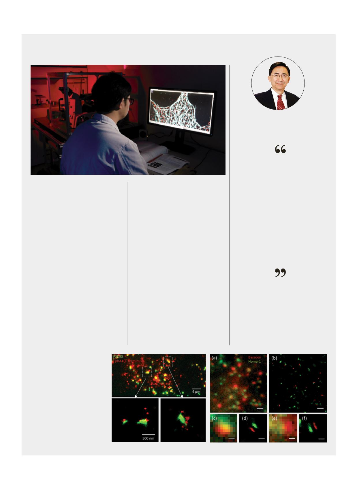
Right: Super-resolved structure of
the Ephrin receptor (EphA4, red) at
peripheral of post synaptic density
(PSD95, green) at synaptic region (Prof
Nancy Ip Lab).
Far right: Super-resolution microscope
can clearly resolve the synaptic
structure in mouse brain. (a)(c) and (e)
show the EPI-fluorescent image which
fails to tell the details of structure. (b)
(d) and (f) are super-resolution images
acquired by a custom-designed two-
color localization microscope. The
pre- and post-synaptic structures are
clearly resolved. Scale bars: 1μm in (a)
and (b); 200nm in (c) and (d).
PROF MICHAEL LOY
Chair Professor of Physics,
Co-director, HKUST Super-Resolution
Imaging Center
The whole system of super-
resolution microscopy is
quite complex. If you are a
biologist, you might not know
what to do with it. If you are
a physicist, you might not
know what to look at. We
recognized this and the two
disciplines got together
Insights into an InfinitesimalWorld
Two-color super-resolution microscope in use.
light sheet microscope for the observation
of living specimens at ultra-high speed,
assisted by equipment project funding from
the Hong Kong Research Grants Council.
Subcellular processes in single cells or
embryos can now be recorded in multicolor
at speeds of up to 500 frames per second.
This provides a new angle to address many
biological questions.


